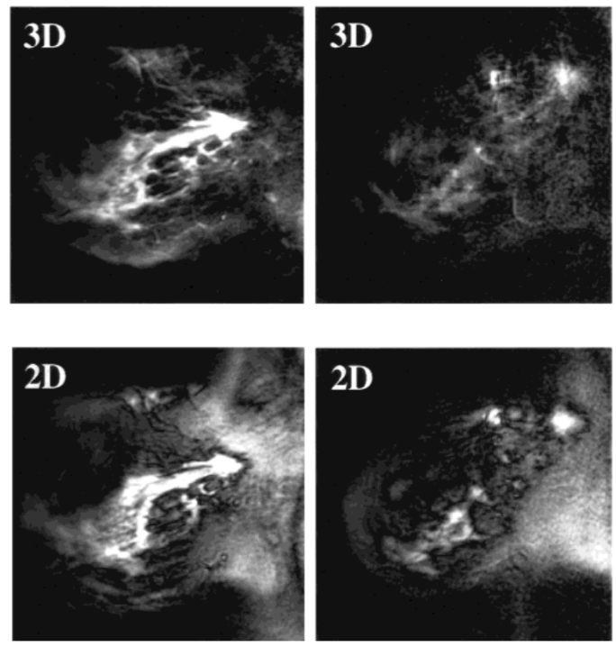
three-dimensional (3)D spiral sequence was developed for dynamic breast magnetic resonance (MR) imaging with much improved image quality. Partial Z phase encoding was applied to obtain thinner slices for a coverage of the whole breast. Comparison between the 3D and a previously developed multi-slice 2D spiral sequences was performed on ten healthy volunteers without contrast and five breast patients with gadolinium-diethylene triamine pentaacetic acid (Gd-DTPA). The 3D spiral images had significantly less off-resonance blurring and spiral artifacts. With a small compromise on temporal resolution (7.7 seconds in 2D and 10.6 seconds in 3D), we obtained 32 interpolated 3-5 mm slices (with 20 Z phase encodes) for a full coverage of 10-16 cm breast with the same 1 x 1 mm2 in-plane resolution as the 2D sequence, which had 12 8-13 mm slices. Contrast between glandular and soft tissue in normal breasts was increased by about 25%. The reduced repetition time in the 3D spiral acquisition led to an increased Gd-enhanced signal. The difference between the enhancement of malignant and benign lesions increased by sevenfold. We expect that this new development could lead to improved specificity in characterizing breast lesions using MR imaging.
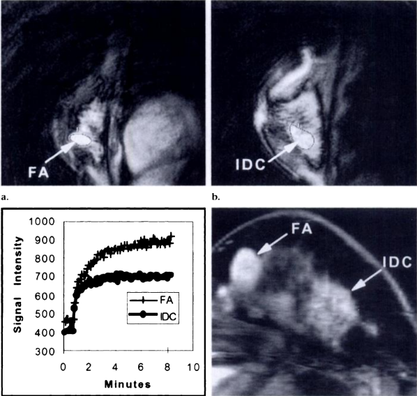
The purpose of this study was to compare various subjective, empiric, and pharmacokinetic methods for interpreting findings at dynamic magnetic resonance (MR) imaging of the breast. Dynamic spiral breast MR imaging was performed in 52 women with, or suspected of having breast disease. Gadolinium-enhanced images were obtained at 12 locations through the whole breast every 7.8 seconds for 8.5 minutes after bolus injection of contrast material. Time-signal intensity curves from regions of interest corresponding to 57 pathologically proved lesions were analyzed by means of a two-compartment pharmacokinetic model, and the diagnostic performance of various parameters was analyzed. Findings included invasive carcinoma in 17 patients, isolated ductal carcinoma in situ (DCIS) in six, and benign lesions in 34. Although some overlap between carcinomas and benign diagnoses was noted for all parameters, receiver operating characteristic analysis indicated that the exchange rate constant had the greatest overall ability to discriminate benign and malignant disease. Dynamic spiral breast MR imaging proved an excellent method with which to collect contrast enhancement data rapidly enough that accurate comparisons can be made between many analytic methods.